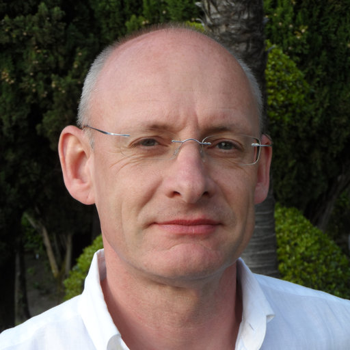
When Oliver Jäkel finished his PhD in theoretical particle physics, he had the ambition to exchange theory for practice in order to have an impact on society. A pilot project to treat cancer patients with light ions fascinated Oliver and made his dream to be a part of it. Through persistence, he made it to the Medical Physics Team of the German Cancer Research Center, where Oliver still works until this day as the Head of the Division of Medical Physics in Radiation Oncology.
At the UT Austin Portugal Program, Oliver is a member of the External Review Committee, a governing body accountable for evaluating the Program and making recommendations to help it stick to its vision and mission and meet the highest scientific standards. Read more about Oliver Jäkel’s story, his views on the progress of cancer research in past years and how he is contributing for it to evolve even further.
How did your professional path cross with cancer treatment, in particular with radiation oncology? How did you decide to embrace the area of Medical Physics?
I did my PhD in theoretical particle physics and, while I finished it, I realized two things: i) my results don’t have any impact on society, as most people would not understand, what I am doing; ii) it is very difficult to make a living in theoretical physics. By chance, I heard about a talk on medical physics as a profession and, at the same time read a lot that the German heavy ion laboratory (GSI) is preparing a pilot project to treat cancer patients with light ions. I was immediately fascinated by this application of particle physics tools and talked to the head of medical physics in the hospital of my university. Amazingly for me, he was quite frustrated about the future perspectives of medical physics, but gave me a contact in Heidelberg at the German Cancer Research Center (DKFZ). Two weeks later I had a postdoc job in the medical physics team, preparing for the patient treatments with carbon ions at GSI. Again, I realized two things: i) it’s always worth trying something, even if chances may be small; ii) if you try, you may be lucky! Today I still work for the DKFZ and the Heidelberg ion beam therapy center of the university.
You are the Head of the Division of Medical Physics in Radiation Oncology of the German Cancer Research Center. Your research focuses on improvements in radiotherapy techniques using photons and ion beams. Can you briefly explain the research your division is currently carrying out?
There are two main areas of our research: a major topic is to introduce adaptive radiotherapy based on high-quality images, e.g. by MRI. This so-called MR-guided therapy gives us the possibility to see the tumor and the surrounding anatomy while we treat. This allows us to react to changes, that we see, be it due to physiological motion (breathing, peristalsis, etc.) or reaction to irradiation (tumor shrinkage, early side effects in normal tissue). Being able to adapt to these changes has a great potential to improve radiotherapy outcomes (e.g. by reducing the treated volume only to the tumor, while in motion) or reduce side effects if we have an image-based prediction already during therapy. There are a lot of tools, we have to develop, in order to make use of all these possibilities clinically, many of them involving advanced methods from computer science, like deep learning and AI. In the field of light ion therapy, there is still also a great potential for improvements, like introducing MR-guidance, but also other possibilities, like using secondary radiation (generated during treatment in the body) for in-vivo monitoring of therapy or the exploration of other ion types, like Helium or oxygen, which have different physical and radio-biological properties.
In your opinion, what are the main challenges related to the research your team is conducting?
The main challenge is to make the step from a development you made in the lab, or your computer and apply it to real-world data, i.e. in the clinic. When applying deep learning methods, a major hurdle is still gathering sufficient structured clinical data for training and validation. The other problem when applying any new development is regulatory issues, like data protection and – even worse – to fulfill the requirements of the medical device regulation. We definitely need dedicated regulations for clinical research!
Some of your projects aim to develop new tools, including algorithms and hardware to support radiotherapy. How do you see the symbiosis between technology and health?
Cancer care, in general, and radiation therapy, in specific, made tremendous progress over the last decades with an incredible improvement in outcomes in many tumors. This is primarily due to the systematic improvement of therapy through advanced technology in both imaging and treatment. With the new treatment techniques, e.g. the MR-Linac or light ion beam therapy, we have a fantastic potential for significant further improvements in many areas of cancer treatment. The exploration of modern technology will continue to improve health care, and the next big step will be applying data science to health. This will be very important, as the demand for cancer therapy is ever-growing due to our ageing societies.
The UT Austin Portugal Program is an example of an international S&T partnership. In your opinion, where is international cooperation most needed when it comes to tackling cancer diseases?
Cancer is not one single disease but a multitude of different diseases with many different aspects. We are already applying tumor genome sequencing and integrating many patient-specific factors to optimize the treatment for individual patients – a concept called personalized therapy. By doing so, we learned that each patient is different and needs a different approach. Being able to address all these different strategies in research and development needs strong international and multi-institutional cooperation on all levels, but especially when it involves designing new concepts for diagnosis and treatment.

