(Closed) TOF-PET for Proton Therapy (TPPT) – In-beam Time-of-Flight (TOF) Positron Emission Tomography (PET) for proton radiation therapy
Summary
The common gamma radiotherapy process lacks accuracy when it comes to analyze prostate, lung, head and neck, liver, esophagus and brain cancers. This project will show the benefits of using TOF-PET in Proton Therapy to increase the performance of Proton Therapy equipment, allowing a more accurate radiation.
The proton therapy used in cancer therapy produces short-lived positron-emitting radioisotopes, such as 15O, 13N and 11C, that can be detected by Positron Emission Tomography (PET) imaging. The strength and time dependence of this image allows assessing the efficacy of the proton treatment in-vivo.
Due to geometrical constraints, the in-beam PET scanning in proton radiation therapy is very difficult to achieve since it is impossible to fully surround the patient with a ring of detectors, as is normally required in PET scanning. However, the use of PET, together with very good TOF, allows obtaining better PET images, with partial angular coverage around the patient.
Once the PET scanner is built, the researchers will use phantom head models to ascertain and verify isotope production maps by a proton beam at MD Anderson. Computer simulations will predict the distribution of the positron annihilation events that should be observed. Any mismatch with the observation will provide feedback to adjust the beam.
The project is going to demonstrate the diagnostic value of the state-of-the-art PET scanner featuring excellent position resolution and Time-of-Flight (TOF) to register positron-emitting radionuclides during and immediately after the proton irradiation.
Expected Outcomes
- A PET system prototype to be used in a Proton Therapy equipment, suitable for radiation monitoring of the head and neck cancers;
- Scientific papers for publication.
| Start Date – End Date: | January 1, 2020 – |
| Scientific Area: | Medical Physics |
| Keywords: | Proton Therapy, TOF PET, Positron Emission Tomography, SiPM readout, TOFPET ASIC |
| Lead Beneficiary (PT): | PETsys Electronics – Medical Pet Detectors, S.A. |
| Co-beneficiaries: | LIP, Laboratório de Instrumentação e Física Experimental de Particulas – Associação para a Investigação e Desenvolvimento Instituto Superior Técnico Universidade de Coimbra |
| PIs at UT Austin: | Karol Lang (College of Natural Sciences, Department of Physics, UT Austin) Narayan Sahoo (Department of Radiation Physics, Division of Radiation Oncology, UT MD Anderson Cancer Center) |
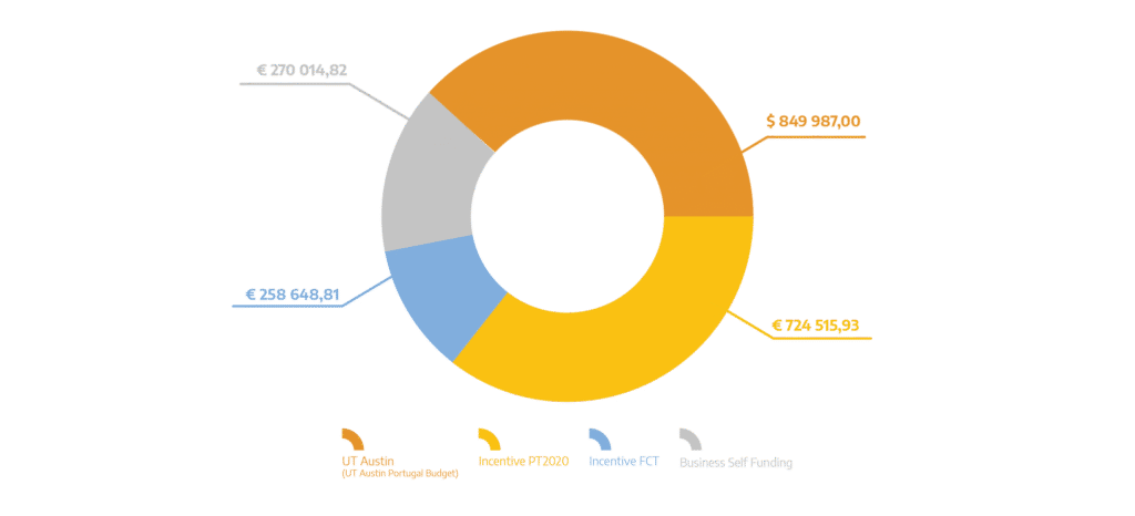
| Total Eligible Investment (PT): | 1 253 179,56 EUR |
| Total Eligible Investment (US): | 849 987,00 USD |
| Funding Sources Distribution: |
Papers and Communications
- Pinto, C. I. G., Guerreiro, J. F., Silva, F., Mendes, F., & Paulo, A. (2023). Radiopharmaceuticals for molecular imaging and theranostics of glioblastoma. In New Insights Into Glioblastoma (pp. 667–705). Elsevier. https://doi.org/10.1016/b978-0-323-99873-4.00023-2
- Abouzahr, F., Cesar, J. P., Crespo, P., Gajda, M., Hu, Z., Kaye, W., Klein, K., Kuo, A. S., Majewski, S., Mawlawi, O., Morozov, A., Ojha, A., Poenisch, F., Polf, J. C., Proga, M., Sahoo, N., Seco, J., Takaoka, T., Tavernier, S., … Lang, K. (2023). The first PET glimpse of a proton FLASH beam. In Physics in Medicine & Biology (Vol. 68, Issue 12, p. 125001). IOP Publishing. https://doi.org/10.1088/1361-6560/acd29e
- Silva, F., Mendes, C., D’Onofrio, A., Campello, M. P. C., Marques, F., Pinheiro, T., Gonçalves, K., Figueiredo, S., Gano, L., Ravera, M., Gabano, E., & Paulo, A. (2023). Image-Guided Nanodelivery of Pt(IV) Prodrugs to GRP-Receptor Positive Tumors. In Nanotheranostics (Vol. 7, Issue 1, pp. 22–40). Ivyspring International Publisher. https://doi.org/10.7150/ntno.78807
- Lang, K. (2022). Towards high sensitivity and high-resolution PET scanners: imaging-guided proton therapy and total body imaging. In Bio-Algorithms and Med-Systems (Vol. 18, Issue 1, pp. 96–106). Walter de Gruyter GmbH. https://doi.org/10.2478/bioal-2022-0079
- Coelho, C. M., Pereira, L., Teubig, P., Santos, P., Mendes, F., Viñals, S., Galaviz, D., & Herrera, F. (2022). Radiation as a Tool against Neurodegeneration—A Potential Treatment for Amyloidosis in the Central Nervous System. In International Journal of Molecular Sciences (Vol. 23, Issue 20, p. 12265). MDPI AG. https://doi.org/10.3390/ijms232012265
- Marques, A., Belchior, A., Silva, F., Marques, F., Campello, M. P. C., Pinheiro, T., Santos, P., Santos, L., Matos, A. P. A., & Paulo, A. (2022). Dose Rate Effects on the Selective Radiosensitization of Prostate Cells by GRPR-Targeted Gold Nanoparticles. In International Journal of Molecular Sciences (Vol. 23, Issue 9, p. 5279). MDPI AG. https://doi.org/10.3390/ijms23095279
- Layden, C., Klein, K., Matava, W., Sadam, A., Abouzahr, F., Proga, M., Majewski, S., Nuyts, J., & Lang, K. (2022). Design and modeling of a high resolution and high sensitivity PET brain scanner with double-ended readout. In Biomedical Physics & Engineering Express (Vol. 8, Issue 2, p. 025011). IOP Publishing. https://doi.org/10.1088/2057-1976/ac4f0a
- Silva, F., D’Onofrio, A., Mendes, C., Pinto, C., Marques, A., Campello, M. P. C., Oliveira, M. C., Raposinho, P., Belchior, A., Di Maria, S., Marques, F., Cruz, C., Carvalho, J., & Paulo, A. (2022). Radiolabeled Gold Nanoseeds Decorated with Substance P Peptides: Synthesis, Characterization and In Vitro Evaluation in Glioblastoma Cellular Models. In International Journal of Molecular Sciences (Vol. 23, Issue 2, p. 617). MDPI AG. https://doi.org/10.3390/ijms23020617
- Cudalbeanu, M., Peitinho, D., Silva, F., Marques, R., Pinheiro, T., Ferreira, A. C., Marques, F., Paulo, A., Soeiro, C. F., Sousa, S. A., Leitão, J. H., Tăbăcaru, A., Avramescu, S. M., Dinica, R. M., & Campello, M. P. C. (2021). Sono-Biosynthesis and Characterization of AuNPs from Danube Delta Nymphaea alba Root Extracts and Their Biological Properties. In Nanomaterials (Vol. 11, Issue 6, p. 1562). MDPI AG. https://doi.org/10.3390/nano11061562
- Pinto, C. I. G., Bucar, S., Alves, V., Fonseca, A., Abrunhosa, A. J., da Silva, C. L., Guerreiro, J. F., & Mendes, F. (2020). Copper-64 Chloride Exhibits Therapeutic Potential in Three-Dimensional Cellular Models of Prostate Cancer. In Frontiers in Molecular Biosciences (Vol. 7). Frontiers Media SA. https://doi.org/10.3389/fmolb.2020.609172
- Silva, F., Paulo, A., Pallier, A., Même, S., Tóth, É., Gano, L., Marques, F., Geraldes, C. F. G. C., Castro, M. M. C. A., Cardoso, A. M., Jurado, A. S., López-Larrubia, P., Lacerda, S., & Cabral Campello, M. P. (2020). Dual Imaging Gold Nanoplatforms for Targeted Radiotheranostics. In Materials (Vol. 13, Issue 3, p. 513). MDPI AG. https://doi.org/10.3390/ma13030513
- Francisco Silva, António Paulo, Agnès Pallier, Sandra Même, Éva Tóth, Lurdes Gano, Fernanda Marques, Carlos FGC Geraldes, M Margarida CA Castro, Ana M Cardoso, Amália S Jurado, Pilar López-Larrubia, Sara Lacerda, Maria Paula Cabral Campello. Dual Imaging Gold Nanoplatforms for Targeted Radiotheranostics. In: José Alexandre Bogas, editor. Prime Archives in Material Science: 2nd Edition. Hyderabad, India: Vide Leaf. 2020.